Sunday 28 May 2017

Fertilization of the Mussels

OBJECTIVES
To differentiate the sexual dimorphism in mussels and to observe the fecundation of them.

MATERIAL 

  • A knife or a cutter 
  • Slide and a coverslip 
  • Tank work 
  • Forceps
  • Dropper
  • Optical microscope 
  • Needles 
  • Distilled water
  • Sea water 
  • Mussels (male and female)

PROCEDURE 

  • Buy a kilo of mussels, they have to be closed and fresh. 
  • Open them with a knife or a cutter, cutting the borders until the two muscles that keep them closed had broken. 
  • The reproductive system of the mussel is formed by gonads, which in the sexually mature individuals occupy the greater part of the body, and especially the mantle. In the females they have a red-orange color more or less intense and in the males white-cream.
  • Break a small piece of the female mantle that it must covered by sea water with the help of a forceps. 
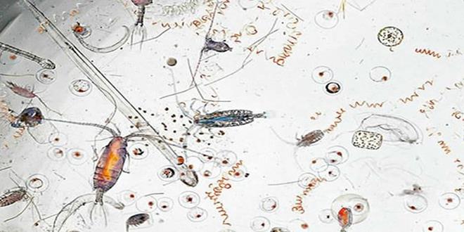
  • With a dropper, take a little part of the pink liquid (ovum) and put them in a slide.
  • Repite the previous operation but now with the male. Take with a dropper the white liquid(sperm)  in the same slide but in a different side of the pink liquid.
  • With the needles establish a bridge between them. And observe in the microscope. 

RESULTS 


QUESTIONS 


  • Differences betweeen male and female mussel
  • The reproductive system of the mussel is formed by gonads, which in the sexually mature individuals occupy the greater part of the body, and especially the mantle. In the females they have a red-orange color more or less intense and in the males white-cream.

  • Mark the anatomical parts of the mussel.


  • Family / class / order / suborder in Latin.
Mytilidae, bivalvia, mytiloida, pteriomorphia. 
  • Do the gametes move? Why the male mussel have the tail smaller than in human esperamtozoides? 
They do not move and the tail of an espermatozoide is smaller because it doesn't have to arrive until the ovum. The two gametes fuse in the middle.
  • Internal or external fertilization?

Ovuliparity is a type of oviparity, a process of sexual reproduction by which both the fertilization of the zygote -union of the male and female gametes- and the development of the embryo occurs in the external environment, outside the urogenital apparatus of the female.

Sunday 14 May 2017

CELL MITOSIS

OBJECTIVES

The aim of this practice is to identify the phases of the mitosis in the root of an onion.

MATERIAL 

  • Microscope
  • Slide and coverslide
  • Dropper
  • Watch glass.
  • Beaker
  • Forceps
  • Filter paper
  • Scissors
  • Bunsen burner
  • Lighter.
  • Needles. 
  • Distilled water
  • Orcein A
  • Orcen B
  • Onions
  • Water

PROCEDURE


  • We keep in a glass the bulb of an onion for a few days, the glass must contain water that is in contact with the roots until the roots measure 3mm.
  • Then, we cut the roots and we put them in a watch glass and we add 2mL of Orcein A.
  • Heat the watch glass with the orcein until we see gray vapors.
  • After that, take the root and put it on the slide and add drops of orcein B. 
  • We place the slide on filter paper, wrapping and tighting it.
  • We cover it with the coverslide and we observe the results on the microscope. 


RESULTS 

If it has worked out well, the process of mitosis must be observed.


EXTRA

Fossilized lizard


Monday 24 April 2017

P20 YOGURT BACTERIA


OBJECTIVES
To reveal bacteria that stay in yogurt such as Streptococcus Thermophilus and Lactobacillus Bulgaricus and to know how to do the Gram Stain in order to reveal them.

MATERIAL
  • Crystal violet
  • Ethanol
  • Distilled water
  • Safranin
  • Three beakers
  • Dropper
  • Bunsen burner
  • Lighter
  • Forceps
  • Slide and coverslide
  • Dissection needle
  • Iodine (Lugol)
  • Petri dish


PROCEDURE

Take the dissection needle and burn it in the Bunsen burner and then spread it on the yogurt.


Put the yogurt on a slide, then take a dropper and throw a few drops of Chrystal violet, wait for 1 minute and 30 seconds.  After that clean the slide with some distilled water.


Now do the same with Iodine, throw on it few drops and wait for 1 minute, then clean it with water again.


Wash it well with ethanol.


Now you have to dye the slide with safranin and to put again some water, after that add the coverslide and put it into the microscope. 


RESULTS 
The bacteria that are observed of violet colour belong to the gram positive (G +), whereas those that are observed of pink colour belong to the gram negative (G-).


Streptococcus Thermophilus and Lactobacillus Bulgaricus belong to G+ because they don't have a light pink colour. 

QUESTIONS

EXPLANATION OF GRAM STAIN

The first step in any staining should always be fixing with heat. Then, by adding the violet crystal, it enters all bacterial cells, both Gram positive and Gram negative).
Lugol is formed by I2 (iodine) in equilibrium with KI (potassium iodide), which is present to solubilise iodine. I2 enters the cells and forms an insoluble complex in aqueous solution with the violet crystal.
The alcohol-acetone mixture which is added serves to effect the discoloration, since the I2 / violet crystal complex is soluble therein. Gram-positive organisms do not discolour, while Gram-negative organisms do. To disclose the Gram negative cells a contrast dye is used. It is usually a red dye, such as safranin. After contrast staining the Gram negative cells appear red, while the Gram positive cells remain blue.


OBSERVATION OF G+ OR G- 

The first membrane belongs to Gram + and the second to Gram -. 


Sunday 2 April 2017

P19 CROMATOGRAPHY


OBJECTIVES

  • To know the bases of chromatographic analysis.
  • To learn the procedure for separating the photosynthetic pigments. 
MATERIAL 
  • ethanol
  • Funnel
  • paper 
  • spatula
  • Cromatography paper
  • mortar
  • Petri dish
  • spinach 
  • Calcium carbonate


PROCEDURE

  • Wash the spinach and cut them, then dry them.
  • Grind the spinach in a mortar and add 50 ml of ethanol and a small amount of calcium carbonate, which fits at the tip of the spatula.
  • Filter the mixture into a funnel with filter paper.
  • Put a part of the mixture in a Petri dish (3-4 mm high).
  • Put a rectangle of paper for chromatography in form V, and wait for the pigments to be differentiated.


RESULTS

The different pigments that are from bottom to top are:
Beta carotene (red - orange)
Chlorophyll alpha (bluish green)
Chlorophyll beta (bright light green)
Xanthophylls (yellow)


QUESTIONS
Why do we add calcium carbonate?
To avoid the degradation of the pigments.
Which is the colour of every pigment
  • Beta carotene (red - orange)
  • Chlorophyll alpha (bluish green)
  •  Chlorophyll beta (bright light green
  • Xanthophylls (yellow)

For what purpose it does have different colour pigments?
To know the different solubility of photosynthetic pigments in an organic solvent such as ethanol, which is miscible with water.


Why do they separate in the paper? 
When the alcohol is placed with the pigments dissolved by a chromatography paper, the different pigments are separated as the medium is increasingly poorer in ethanol and richer in water.

Monday 27 March 2017

P18 WATER DROP

OBECTIVES
The aim of this practice is to observe water from a fishbowl and from a river and to know that there is life in the environment of water. 


MATERIAL 
  • Slide and a coverslide.
  • Water from a fishbowl.
  • Water from a river. 
  • Microscope.
  • A dropper.

PROCEDURE

  • Prepare two samples, one with the water of the river and the other the water was from the fishbowl. 
  • So,take one drop from each type of water and put add them in a slide, then cover them with the coverslide.
  • Put them in the corresponding microscope and observe the results.


CONCLUSIONS 
  • In the water from the river we saw arthropods.
Exemple water of the river. 


  • In the water of the fishbowl we observe protozoans. 
Exemple water of the fishbowl.


Saturday 11 March 2017

P17 AMYLOPLASTS AND CHROMOPLASTS

OBJECTIVES

The aim of this practice is to observe cells from tomato, carrot and potato to identify amyloplasts and chromoplasts.

MATERIAL
  • A potato
  • A carrot
  • A tomato
  • A cutter or a knife
  • A slide and a coverslide
  • Distilled water
  • An optical microscope
  • A dropper
  • A forceps


PROCEDURE

TOMATO
  • Make a thin cut in the tomato pulp of 1 or 2 mm.
  • Place the cut on the slide without adding water.
  • Then cover with coverslip and crush gently.
  • Observe in the optical microscope.

CARROT

Do the same as with the tomato but adding distilled water to be able to observe the sample well.

POTATO

A

Cut a piece of the epidermis from the potato and observe it under an optical microscope.

B
  • Take a few drops of potato whey with the help of a dropper.
  • Then place the droplets on the slide and cover with the coverslip.
  • We observe it under a microscope.


RESULTS 

Chromoplasts of the tomato


Chromoplasts of the carrot 


Amiloplasts of the potato (serum and epidermis)


CONCLUSIONS

The plast are vacuoles that carry the production and store of nutrients and plant cells.

The chromoplasts contain pigments that bring the coloration to the vegetable.

The leucoplasts contain substances of store like starch, with are called amyloplasts.


Monday 27 February 2017

P16 C.ELEGANS

INTRODUCTION 

On the 15th of February we went to the PRBB, biomedical research park in Barcelona. There we did an activity that consisted in classifying some bugs (some worms called C. Elegans) according to the characteristics that they had (physical and genetic).

There were 4 small boxes:

- Wild C. elegans.
- GFP + RFP (fluorescence proteins)
- Daf 18 mutated (mutated gene, they had cut it so that C. Elegan was shorter).
- Daf 18 silenced (turn off a gene).

We divided into four groups (Microscopy, image, genetics and behaviour). One of our students went through the groups analysing what we were doing.

My group had the tasks of microscopy.

OBJECTIVES 

The aim of microscopy was to classify the C. Elegans from knowing that in the laboratories there are more hermaphrodites, so that they can reproduce better. Hermaphrodites have a pointed tail and the males have a round tail. 

PROCEDURE 

We take some microscopes and one magnifying glass to see the different boxes of c. elegans and we keep watching them and to check how many were males and hermaphrodites. We took notes in a paper. 


RESULTS 
These are the common results of all groups.


TYPE (RESULT)
MICROSCOPY
IMAGE
GENETICS
BEHAVIOUR
A
DAF 18 mutated
(FLUORESCENCE)
8 males
100 herma
I X
F X
More shorter
More to the salt. (THEY SMELL)
B
GFP-RFP

All herma
I X
F V
Long
More to the salt.(THEY SMELL)
C
WILD
6 males
100 herma
I X
F X
Long
They go wherever they want.( THEY DON’T)
D
DAF 18 SILENCED
All herma

I X
F X
Less long than the others
They go wherever they want. (THEY DON’T)


P15 OSMOSIS IN THE ONION


OBJECTIVE 


The aim of this practice is to observe the process of osmosis in the onion, as well as to see the plasmolysis and the turgor  of the plant cell in each case using salt and distilled water.

MATERIAL 
  • Optical microscope 
  • A slide and a cover slide
  • Forceps
  • A dropper  
  • An onion 
  • A knife 
  • Distilled water
  • Salt
  • A  100mL beaker
PROCEDURE 

Peel the onion and cut two small pieces of the inside.

Prepare two optical microscopes.
Prepare a beaker with water and salt.
Make a sample with one of the pieces and add a few drops of salt water with the help of a dropper.
Cover it with the slide and coverslip, and watch it. In this case it is not necessary to dye it.


Then prepare another sample but this time adding drops of distilled water.
Cover and observe under a microscope.


RESULTS
The sample with salt is called hypertonic solution, so the cells are going to lose water and became smaller and tighter.


The sample with distilled water is an isotonic solution, so the cells are going to absorb water and became bigger and fatter. 


QUESTIONS 


   1. When the salt solution was added to the onion cells where was the greater concentration of water? The higher concentration of water will be outside of the cell, because of the hypertonic solution, the water will go outside to equal the concentration.

   2. Which kind of transport does water follow across the membrane? Active transport by simple diffusion.